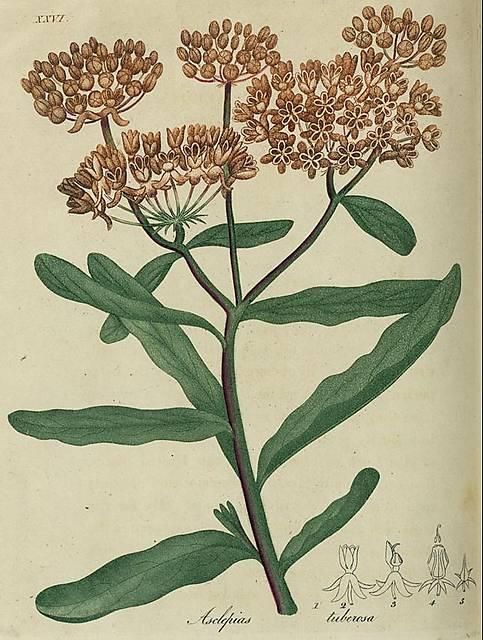
Plate XXI.
Fig. 1. Asclepias tuberosa.
Fig. 2. A flower.
Fig. 3. A flower dissected, showing the mass of anthers, and one nectary with its horn.
Fig. 4. Magnified section of the mass of anthers, showing the situation of the pistils inside, &c. A pair of pollen masses is drawn out at the top.
Fig. 5. Pistils magnified, and calyx.
This image is from Asclepias in American Medical Botany, Bigelow, 1817-1821.
(used in Rafinesque)


