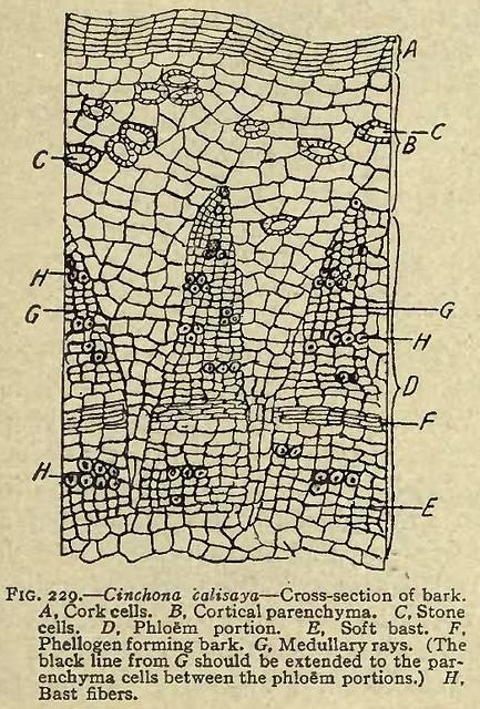
A, Cork cells. B, Cortical parenchyma. C, Stone cells. D, Phloem portion. E, Soft bast. F, Phellogen forming bark. G, Medullary rays. (The black line from G should be extended to the parenchyma cells between the phloem portions.) H, Bast fibers.
This image is from Cinchona in Sayre's Manual of Organic Materia Medica and Pharmacognosy, 1917.


