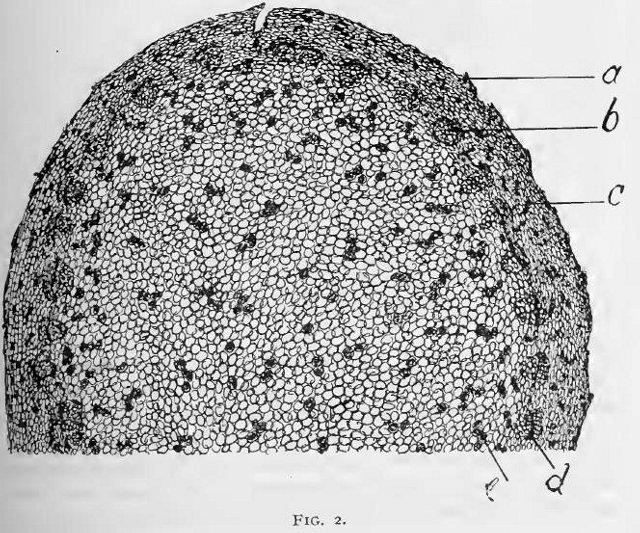
magnified 15 diameters.
a, cork;
b, vasal bundle;
c, cluster of secretion-cells in middle bark;
d, cambium;
e, secretion-cells in pith.
This image is from Some further observations on the structure of Sanguinaria canadensis in the American Journal of Pharmacy, 1895.


