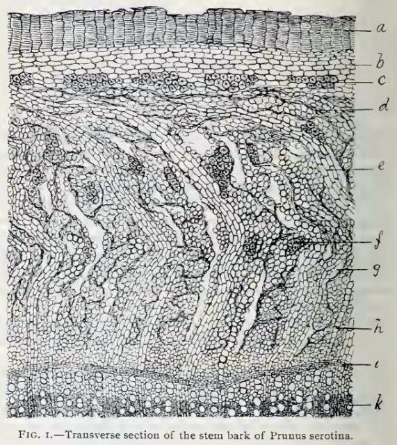
Fig. 1.—Transverse section of bark of Prunus serotina magnified 75 diameters. The specimen was from a stem only five or six years old.
a, cork, probably secondary periderm;
b, middle or green layer of bark;
c, clusters of stone cells in inner portion of middle bark;
d, compressed sieve tissue in the outer portion of bast layer;
e, a medullary ray;
f, mass of stone cells;
g, fissure between medullary ray and bast;
h, medullary ray;
i, cambium zone;
k, duct in mature wood.
This image is from Structure of our Cherry Barks in the September issue of the American Journal of Pharmacy, 1895.


