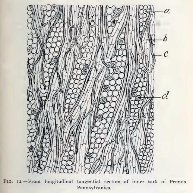
Fig. 12.—Portion of longitudinal tangential section of inner bark of P. Pennsylvanica magnified about 75 diameters.
a, medullary ray;
b, soft bast cell;
c, bast fibre;
d, crystal cell.
This image is from Structure of our Cherry Barks in the September issue of the American Journal of Pharmacy, 1895.


