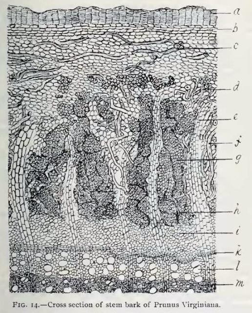
Fig. 14.—Cross section of stem bark of P. Virginiana, magnified about 75 diameters.
a, periderm;
b, outer cortex (collenchyma);
c,d, tortuous sclerenchyma fibres;
e, medullary ray;
f, sclerenchyma fibre;
g, large mass of secondary bast fibres;
h, compressed sieve tissues separating masses of bast fibres;
i, younger bast;
k, cambium zone;
l, duct in newly formed wood;
m, mature wood.
This image is from Structure of our Cherry Barks in the September issue of the American Journal of Pharmacy, 1895.


